Antipodal symmetry and asymmetry in the embryo sac of Avena sativa L.
Abstract
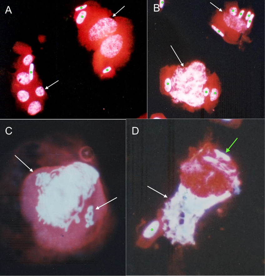
Inter- and intra-antipodal variation is presented for a free-nucleolar developmental stadium in the Avena sativa endosperm. Antipodals of common oat are uni- or multinuclear. Multinuclear antipodals, in a single cell, show nuclei of the same mitotic stage, but this is different for various antipodals. Examples of DNA amplification and anomalies occurring during mitosis are provided.
References
Batygina T.B. 1987. Chlebnoe zerno. Nauka, Leningrad.
Chaban I.A., Lazareva E.M., Kononenko N.V., Polyakov V.Y. 2011. Antipodal complex development in the embryo sac of wheat. Russ. J. Develop. Biol. 42: 79–91.
Ivanovskaya E.V. 1983. Citoèmbriologičeskoe issledovanie differencirovki kletok rastenij. Izdatel’stvo Moskovskogo Universiteta, Moskva.
Kosina R. 1994. Cytogenetyka molekularna amfiploidów: tetraploidy Triticum × Aegilops squarrosa. Sprawozdania Wrocławskiego Towarzystwa Naukowego 49B: 57–60.
Krawczyk J. 2008. Zmienność cytogenetyczna bielma u wybranych heksaploidów Avena L. MSc thesis. University of Wroclaw, Wroclaw.
Schwarzacher T., Ambros P., Schweizer D. 1980. Application of Giemsa banding to orchid karyotype analysis. Plant Syst. Evol. 134: 293–297.
Wędzony M. 1992-1993. Polytenization in the antipodal nuclei of wheat [Triticum aestivum L.], triticale [×Triticosecale Wittm.] and their reciprocal crosses. Acta Biol. Cracov. Ser. Bot. 34-35: 43–57.

This work is licensed under a Creative Commons Attribution-NonCommercial-NoDerivatives 4.0 International License.
The journal is licensed by Creative Commons under BY-NC-ND license. You are welcome and free to share (copy and redistribute the material in any medium or format) all the published materials. You may not use the material for commercial purposes. You must give appropriate credit to all published materials.
The journal allow the author(s) to hold the copyrights and to retain publishing rights without any restrictions. This is also indicated at the bottom of each article.