Structure and chemical content of the trichomes in two Doronicum species (Asteraceae)
Abstract
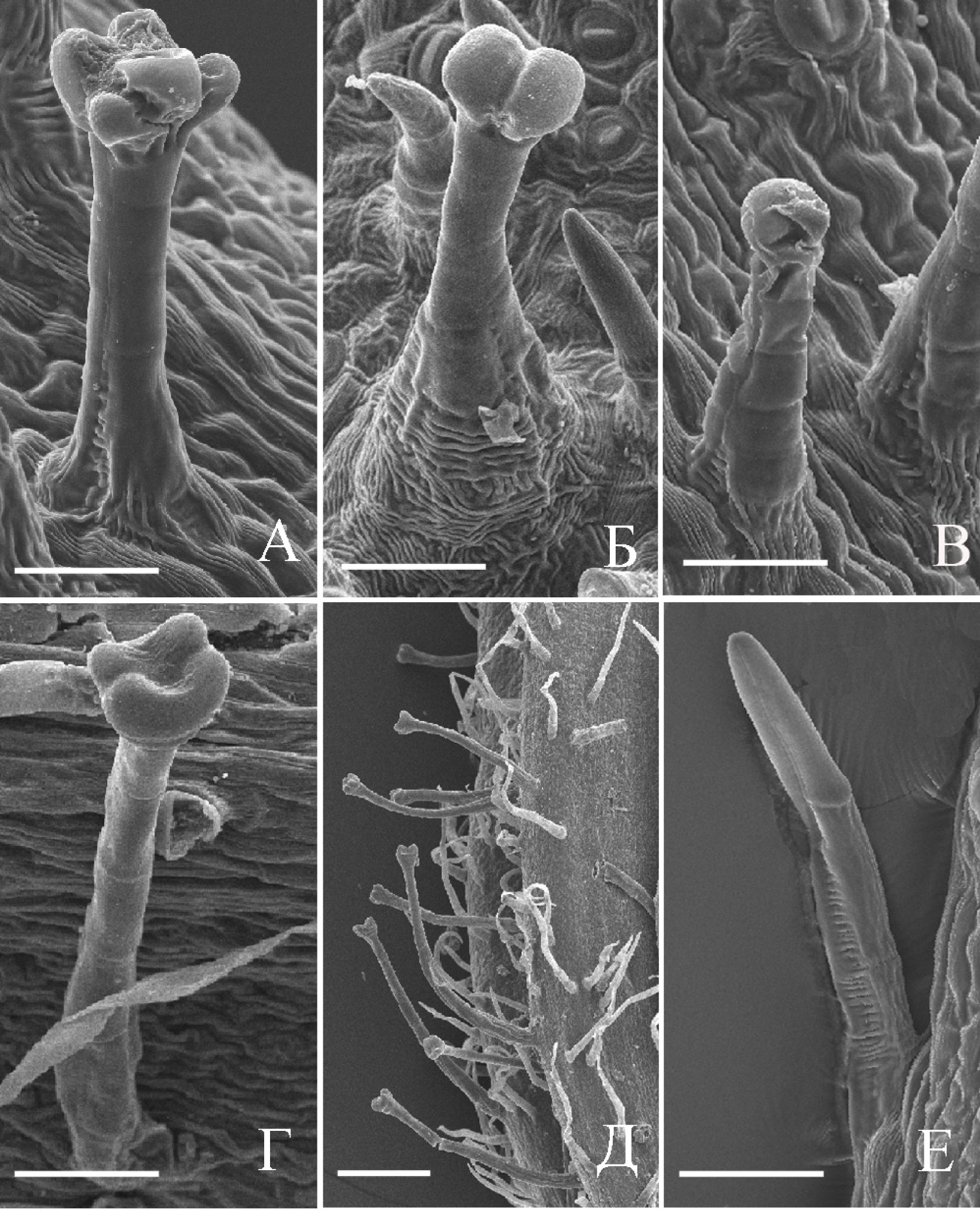
Non-glandular and three types of glandular trichomes localized on a peduncle and the phyllaries of two species (Doronicum orientale and D. macrophyllum) belonging to Senecioneae tribe of Asteraceae were studied by light and electron microscopy, as well as on histochemistry. The plant species differ on the trichome morphology, anatomy and arrangement. Three types of the glandular trichomes are formed on the vegetative and reproductive organs in D. orientale, whereas only one type exists in D. macrophyllum. It was shown by fluorescent microscopy and histochemistry that the phenolic substances, terpenoids and sesquiterpene lactones present in the glandular trichomes. The received results allow assuming that secretion of the trichomes is a chemical barrier to insects and fungi.
References
Adedeji O. 2008. Importance of leaf epidermal characters in the Asteraceae family. Not. Bot. Hort. Agrobot. Cluj 36 (2): 7–16.
Andreucci A., Ciccarelli D., Desider M., Pagni M. 2008. Glandular hairs and secretory ducts of Matricaria (Asteraceae): morphology and histochemistry. Ann. Bot. Fennici 45: 11–18.
Appezzato-da-Gloria B., Costa F.B., Silva V.C., Gobbo-Neto L., Rehder V.L.G., Hayashi A.H.H. 2012. Glandular trichomes on aerial and underground organs in Chrysolaena species (Vernonieae – Asteraceae): structure, ultrastructure and chemical composition. Flora 207 (12): 878–887.
Ascensao L., Pais M.S. 1982. Secretory trichomes from Artemisia crithmifolia: some ultrastructural aspects. Bull. Soc. Bot. Fr., Act. Bot. 129 (1): 3–7.
Cappelletti E.M., Caniato R., Appendino G. 1986. Localization of the cytotoxichydroperoxyeudesmanolides in Artemisia umbelliformis. Biochem. Syst. Ecol. 14: 183–190.
Ciccarelli D., Garbari F., Pagni A. 2007. Glandular hairs of the ovary: a helpful character for Asteroideae (Asteraceae) taxonomy? Ann. Bot. Fenn. 44: 1–7.
Combrink S., Plooy G.W., McCrindle R.I., Botha B.M. 2007. Morphology and histochemistry of the glandular trichomes of Lippia scaberrima (Verbenaceae). Ann. Bot. 99: 1111–1119.
Corsi G., Bottega S. 1999. Glandular hairs of Salvia officinalis: new data on morphology, localization and histochemistry in relation to function. Ann. Bot. 84: 657–664.
Cron G., Balkwill K., Knox E. 2006. Two new species of Cineraria (Senecioneae, Asteraceae) from KwaZulu‑Natal, South Africa. S. Afr. J. Bot. 72: 34–39.
David R., Carde J-P. 1964. Coloration differentiele des inclusions lipidique et terpeniques des pseudophilles du pin maritime au moyen du reactif nadi. C. R. Acad. Sci. 258: 1338–1340.
Duke S., Paul R. 1993. Development and fine structure of the glandular trichomes of Artemisia annua L. Int. J. Plant Sci. 154 (1): 107–118.
Geissmann T.A., Griffin T.S. 1971. Sesquiterpene lactones: acid-catalysedcolor reactions as an aid in structure determination. Phytochem. 10: 2475–2485.
Gutmann M. 1995. Improved staining procedures for photographic documentation of phenolic deposits in semi-thinsections of plant tissue. J. Microsc. 179: 277–281.
Heinrich G., Pfeifhofer H., Stabentheiner E., Sawidis T. 2002. Glandular hairs of Sigesbeckia jorullensis Kunth (Asteraceae): morphology, histochemistry and composition of essential oil. Ann. Bot. 89: 459–469.
Metcalfe C.R., Chalk L. 1950. Anatomy of the decotyledons. 2nd ed. Clarendon Press, Oxford.
Nikolakaki A., Christodoulakis N. 2004. Leaf structure and cytochemical investigation of secretory tissues in Inula viscosa. Bot. J. Linn. Soc. 144: 437–448.
Pagni A.M. 1995. Secretory structures in the capitula of Santolina leucantha Bertol. (Asteraceae). Morphology and histochemistry. Ann. Bot. (Roma) 53: 239–249.
Pagni A.M., Orlando R., Masini A., Ciccarelli D. 2003. Secretory structures of Santolina ligustica Arrigoni (Asteraceae) an Italian endemic species. Israel J. Plant Sci. 51: 185–192.
Swenson U. 1995. Systematics of Abrotanella, an amphi-pacific genus of Asteraceae (Senecioneae). Pl. Syst. Evol. 197: 149–193.

This work is licensed under a Creative Commons Attribution-NonCommercial-NoDerivatives 4.0 International License.
The journal is licensed by Creative Commons under BY-NC-ND license. You are welcome and free to share (copy and redistribute the material in any medium or format) all the published materials. You may not use the material for commercial purposes. You must give appropriate credit to all published materials.
The journal allow the author(s) to hold the copyrights and to retain publishing rights without any restrictions. This is also indicated at the bottom of each article.