Anatomical structure of Trapa natans L. submerged organs in context of the species ecology
Abstract
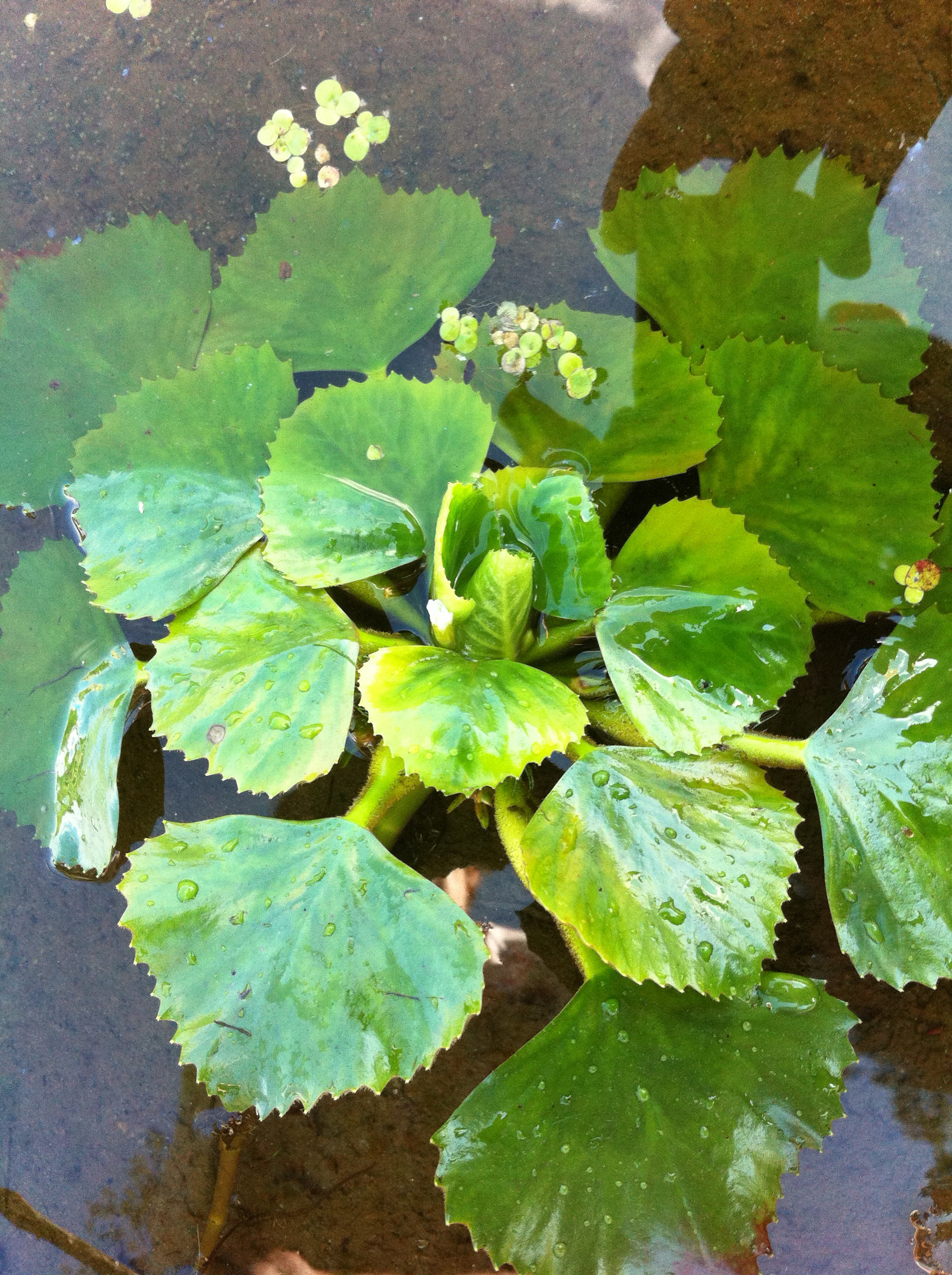
The results of anatomical study of submerged organs of Trapa natans (water chestnut) in budding-flowering stage are presented. The plants were collected along the shore of the Rusanivsky channel (left shore of Dnepr River, in Kiev). The light and laser scanning confocal microscopy were used. Anatomical structure of the submerged secondary roots, the submerged dissected leaves and stem had the signs that are typical for submerged organs of some hydrophytes. But, the roots are characterized by the peculiarity, they were photosynthesizing. Chloroplasts are revealed in root core cells. The middle number of chloroplasts in one parenchyma cell of submerged leaf was four times more than in one core cell of root. The fluorescence intensity of leaf chloroplasts was also more than that in root or stem chloroplasts. The very developed aerenchyma was characterized for roots and submerged stem in comparison with the submerged dissected leaves, in which an aerenchyma was weak developed and it was presented by very small air space between the cells of inner layer in parenchyma.
References
Езау К. 1980. Анатомия растений. Кн. 2. Мир, Москва.
Медведев С.С. 2004. Физиология растений. Изд. Санкт-Петербургского ун-та, Санкт-Петербург.
Недуха О.М. 2011. Гетерофілія у рослин. Альтерпрес, Київ.
Недуха О.М., Котенко Т.Б. 2012. Гетерофілія у Trapa natans L. Морфолого-анатомічна будова листків. Mod. Phytomorphol. 2: 29–33.
Ронжина Д.А., Пьянков В.И. 2001. Структура фотосинтетического аппарата листа пресноводных гидрофитов. ІІ. Количественная характеристика мезофилла листа и функциональная активность листьев с разной степеню погружения. Физиология растений 48: 836–845.
Фурст Г. 1979. Методы анатомо-гистохимического исследования растительных тканей. Наука, Москва.
Arber A. 2008. Water plants. A study of aquatic angiosperms. Cambridge University Press, Cambridge.
Armstrong W., Brande R., Jackson M.B. 1994. Mechanisms of flood tolerance in plants. Acta Bot. Neerland. 43: 307–358.
ARS 2004. Agricultural Research Service. Systematic botany and mycology laboratory. United States, NY. http://www.ars.usda.gov/main/main.htm
Bercu R. 2004. Histoanatomy of the leaves of Trapa natans (Trapaceae). Phytologia Balcanica 10: 51–55.
Bercu R. 2010. Histoanatomy of Barcopa carolina (Walt) Robins. (Scrophulariaceae). Biologie (Universitatea”Vasile Alecsandri” din Bacău) 18: 99–102.
Bercu R., Fagaras M. 2002. Anatomical features of the root, stem and leaf blade of Potamogeton pectinatus L. and Vallisneria spiralis L. Conf. Bot. Univ “Babes-Bolyal” Cluj-Napoca 37: 41–42.
Bitonti M., Cozza R. , Wang G., Ruffini-Castiglione M., Mazzuca S., Castiglione S., Sala F., Innocenti A.M. 1996. Nuclear and genomic changes in floating and submerged buds and leaves of heterophyllous water chestnut (Trapa natans). Physiol. Plantarum 97: 21–27.
Crawford R.M.M. 1983. Root survival in flooded soils. In: Gore A.J.P. (ed.), Ecosystems of the world, 4A, Milers: swamp, bog, fen and moor: 257–283. Elsevir Sci. Publ. Co., Amsterdam.
Currier H.B., Shin C.Y. 1968. Sieve tubes and callose in Elodea leaves. Amer. J. Bot. 55: 145–152.
He J.B., Bogemann G.M., Van de Steeg H.G., Rijnders J.G.H-M. 1999. Survival tactics of Ranunculus species in river flood plants. Oecologia 118: 1–8.
Herth W. 1980. Calcofluor white and congo-red inhibit chitin microfibril assembly of Poteriochromonas: evidence for a gap between polymerization and microfibril formation. J. Cell Biology 84: 642–658.
Ishimaru K., Kubota F., Saitou K, Nakayama M. 1996. Photosynthetic response and carboxylation activity of enzymes in leaves and roots of water chestnut, Trapa bispinosa Roxb. J. Fac. Agr., Kyushu Univ. 41: 57–65.
Pierobon E., Bolpagni R., Bartoli M., Viaroli P. 2010. Net primary production and seasonal СО2 and CH4 fluxes in a Trapa natans L. meadow. J. Limnol. 69: 225–234.
Sculthorpe C.D. 1971. The biology of aquatic vascular plants. Edward Arnold, London.
Shalabh B., Akash J., Chaudhary J. 2012. Trapa natans (water chestnut): an overview. Int. Res. J. Pharm. 3: 31–33.
Vartapetian B., Jackson M.B. 1997. Plant adaptation to anaerobic stress. Ann. Bot. 79: 3–20.
Zimmermann J.L. 1993. Cheyenne bottoms. Wetland in Jeopardy. University Press of Kansas, Kansas.

This work is licensed under a Creative Commons Attribution-NonCommercial-NoDerivatives 4.0 International License.
The journal is licensed by Creative Commons under BY-NC-ND license. You are welcome and free to share (copy and redistribute the material in any medium or format) all the published materials. You may not use the material for commercial purposes. You must give appropriate credit to all published materials.
The journal allow the author(s) to hold the copyrights and to retain publishing rights without any restrictions. This is also indicated at the bottom of each article.